Advances in Nash Hcc Screening
Nash HCC screening is a critical element in detecting hepatocellular carcinoma (HCC) at its earliest stages, especially in patients with nonalcoholic steatohepatitis (NASH). This article delves into the significance of NASH HCC screening and its methodologies, focusing on the latest advancements and protocols that have improved early detection and treatment outcomes. With a growing prevalence, understanding these screening practices is more crucial than ever.

Understanding Nash HCC Screening
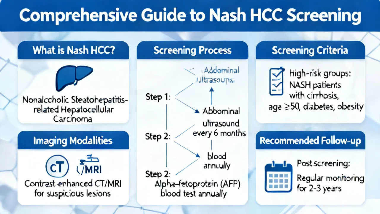
Nonalcoholic steatohepatitis (NASH) is an aggressive form of liver disease characterized by inflammation and damage caused by fat accumulation in the liver. It represents an advanced stage of nonalcoholic fatty liver disease (NAFLD) and can progress to hepatocellular carcinoma (HCC), a primary liver cancer. The increasing prevalence of NASH, correlated with the obesity epidemic and rising diabetes rates, has highlighted its importance in public health. As NASH significantly elevates the risk of HCC, early and precise screening becomes paramount in improving patient outcomes. Modern advances in medical research have introduced more nuanced and effective screening methodologies, allowing healthcare professionals to detect and manage liver cancer in its nascent stages, leading to a more favorable prognosis.
The Importance of Early Detection
Screening for HCC in patients with NASH is crucial as early-stage HCC often presents no symptoms, making it difficult to diagnose without targeted screening efforts. The asymptomatic nature of early HCC means that many patients remain undiagnosed until the disease reaches an advanced, less treatable stage. Therefore, the implementation of effective screening strategies is vital for timely intervention. Timely detection can dramatically improve survival rates and broaden the spectrum of available treatment options. When HCC is detected at an early stage, patients have a significantly higher chance of undergoing successful treatments, such as surgical resection or liver transplantation.
Moreover, early detection allows for better management of both NASH and HCC, considering that liver conditions often coexist. By controlling NASH through lifestyle changes or pharmacotherapy, healthcare providers can potentially slow the progression of liver disease and mitigate the risk of HCC development. This multifaceted approach emphasizes the importance of comprehensive management in high-risk patients.
Current Screening Techniques
The most widely utilized methods for screening NASH-related HCC include ultrasound imaging, blood tests for alpha-fetoprotein (AFP) levels, and magnetic resonance imaging (MRI). Each technique has its strengths and limitations, affecting sensitivity and specificity differently. The choice of a particular method often depends on the patient’s overall health profile, specific risk factors for liver cancer, and available resources. For instance, some patients may have limited access to certain advanced imaging techniques due to cost or availability, thus leaning towards more accessible methods like ultrasound.
Ultrasound Imaging
Ultrasound is the standard first-line screening method due to its non-invasiveness, cost-effectiveness, and wide availability. It operates by using sound waves to create real-time imaging of the liver, helping identify abnormalities that may indicate the presence of HCC. One major advantage of ultrasound is that it does not involve exposure to ionizing radiation, making it safer for routine screenings. However, in the context of NASH, ultrasound may have limited sensitivity in detecting early tumors and smaller lesions, necessitating complementary methods for definitive diagnosis.
In practice, ultrasounds can miss small HCC lesions, particularly those that are less than 1 cm in size, which represents a significant concern for patients with underlying liver conditions such as NASH. To bolster outcomes, healthcare providers often recommend follow-up screenings incorporating additional diagnostic tools, especially when ultrasound results are inconclusive. Additionally, the effectiveness of ultrasound can vary based on patient factors such as body habitus, which may hinder the quality of imaging.
Blood Tests for Biomarkers
Regular blood tests measuring levels of alpha-fetoprotein (AFP) serve as an additional layer of screening for HCC. Elevated AFP levels can indicate liver cancer; however, interpreting these results can be complicated. False positives may occur due to liver inflammation, cirrhosis, or conditions unrelated to cancer, challenging the reliance exclusively on AFP levels for diagnosis.
As a consequence, healthcare providers often use AFP levels in conjunction with imaging techniques for a more comprehensive evaluation. Some studies suggest combining AFP levels with other biomarkers may improve the predictability of liver cancers, facilitating earlier intervention and better patient outcomes. Emerging blood tests, such as des-gamma-carboxy prothrombin (DCP), are also being studied as potential adjuncts to enhance the accuracy of liver cancer screening. These tests collectively contribute to a more nuanced understanding of a patient’s risk of progressing to HCC.
Innovative MRI Approaches
Magnetic Resonance Imaging (MRI), particularly contrast-enhanced MRI, provides detailed imaging that increases accuracy in differentiating between benign and malignant liver lesions. MRI is particularly effective for high-risk patients with inconclusive ultrasound scans or abnormal AFP levels. The high-resolution images generated by MRI allow for a sophisticated assessment of liver architecture and help healthcare providers identify small lesions that may be missed by other methods.
This technique employs contrast agents that enhance the visibility of blood flow and tissue perfusion, thus improving the detection of tumors. Furthermore, MRI's ability to provide functional imaging, through techniques such as diffusion-weighted imaging or MR elastography, enables clinicians to evaluate the cellular environment of liver lesions and offer insights into their potential malignancy.
Despite these advantages, MRI comes with limitations, including higher costs compared to ultrasound and the need for patient compliance, as some individuals may have conditions that prevent them from undergoing MRI, such as claustrophobia or the presence of incompatible medical devices. Additionally, the interpretation of MRI results requires specialized radiological expertise, which may not be accessible in all healthcare settings.
Latest Advances in Screening Technology
Advancements in technology have refined screening practices, enhancing both accuracy and patient experience. Emerging modalities such as elastography—a technique assessing liver stiffness—can detect fibrosis early, offering a predictive marker for liver disease progression and HCC risk in NASH patients. Transient elastography is a non-invasive method gaining traction that allows measurement of liver stiffness, enabling clinicians to stratify patients based on fibrosis stages without the need for liver biopsy.
Elastography can play a pivotal role in patient management by categorizing risk levels, guiding treatment decisions, and monitoring disease progression over time. This sectioning supports healthcare providers in determining the most appropriate resources and interventions, potentially preventing the transition from NASH to HCC.
Furthermore, researchers are actively exploring additional biomarkers and imaging technologies that could further enhance screening protocols. For instance, artificial intelligence is on the horizon, with algorithms being developed to analyze imaging and pathology more effectively. This integration could herald higher detection rates of early-stage HCC by correlating data from various sources, including medical history, genetic factors, and radiologic findings.
Genetic and Molecular Screening Innovations
Genomic and proteomic studies are ushering in a new era in HCC screening, allowing for more personalized and precise evaluations of cancer risk. These advancements enhance early detection and help tailor treatment strategies to individual patients’ molecular profiles, promising improved treatment efficacy.
Recent research is focusing on identifying specific gene mutations or epigenetic changes that may predispose individuals with NASH to develop HCC. Genetic testing can provide essential insights, guiding clinicians in making informed decisions about patient management and screening intervals. Additionally, ongoing pharmacogenomics research is aimed at assessing how patients metabolize drugs differently based on their genetic makeup, ultimately contributing to more personalized treatment plans.
The integration of molecular screening techniques into routine clinical practice could represent a transformative step forward in liver cancer prevention. By identifying high-risk individuals using genetic markers, clinicians can implement proactive management strategies to delay or prevent disease progression while providing comprehensive patient education about lifestyle modifications and other risk reductions.
| Screening Method | Description | Benefits | Limitations |
|---|---|---|---|
| Ultrasound Imaging | Measures liver abnormalities via sound waves. | Non-invasive, cost-effective, and widely available. | Lower sensitivity in detecting small tumors; operator-dependent. |
| Blood Tests (AFP) | Measures alpha-fetoprotein levels in the bloodstream. | Simple, quick, and can be done in outpatient settings. | May produce false positives and not always indicative of liver cancer. |
| MRI | High-resolution imaging using magnetic resonance technology. | High accuracy and detailed visualization of liver lesions. | Costly, may require sedation for claustrophobic patients, and needs expert interpretation. |
| Elastography | Assesses liver stiffness to indicate fibrosis stages. | Predictive marker for disease progression and non-invasive. | Less available in standard clinical settings and can require specific training for use. |
Screening Recommendations and Guidelines
Current guidelines recommend biannual screenings for patients diagnosed with NASH due to their elevated risk for HCC development. Organizations such as the American Association for the Study of Liver Diseases (AASLD) and the European Association for the Study of the Liver (EASL) endorse regular surveillance approaches that combine imaging with serum biomarker testing. A multidisciplinary approach that combines imaging, blood tests, and innovative techniques tends to provide the most reliable detection, aiding in accurate assessments and timely treatments.
In clinical practice, these recommendations emphasize the importance of tailored screening protocols based on individual risk factors. Patients with more severe liver disease or additional risk factors—such as advanced age, male gender, or a family history of liver cancer—may require more frequent monitoring or more aggressive screening techniques. Continuous clinical education and updates to guidelines are essential to keeping healthcare professionals informed about evolving best practices in screening.
Challenges in Implementing Effective Screening
Despite the advancements in screening technologies, several challenges persist in the widespread adoption of effective NASH HCC screening. Access to specialized technologies can be limited in some regions, affecting the equity of healthcare provisions for high-risk populations. Healthcare costs associated with advanced imaging and blood tests can create financial barriers, limiting access to necessary screenings for uninsured or underinsured patients.
Moreover, awareness among both patients and providers about the necessity and benefits of regular screenings remains inadequate. Many at-risk individuals may not recognize their susceptibility to liver disease or may not receive proper counseling regarding their screening options. This highlights the need for public education campaigns to raise awareness about NASH, its risks, and the importance of HCC screening.
Further, achieving a consensus among healthcare providers regarding screening recommendations may prove challenging, particularly as the field of liver disease continues to evolve with new research findings. Clinicians need robust support and resources to navigate the nuances of patient management and to ensure timely and effective screening practices are implemented across diverse healthcare settings.
Future Perspectives
The future of NASH HCC screening appears promising, with ongoing research focused on various dimensions of liver cancer detection and management. The integration of artificial intelligence into diagnostic workflows is one significant area of exploration. Machine learning algorithms could potentially transform the landscape by processing vast patient datasets to predict cancer risks more precisely. These intelligent systems may also assist radiologists in interpreting imaging results more accurately, ultimately reducing the likelihood of misdiagnosis.
Additionally, the development of more specific and sensitive biomarkers for HCC through proteomic and metabolomic studies is underway, offering further potential for routine screening applications. Tests that can assess multiple markers in concert, rather than relying on a single biomarker, may provide a more reliable means of detecting early-stage liver cancer.
As research continues to emerge in this field, the hope is to achieve a comprehensive framework for HCC screening that encompasses early detection, personalized risk assessments, and effective treatment strategies tailored to the needs of individual patients. This holistic approach aligns with the evolving understanding of NASH as a complex disease requiring multidimensional management and underscores the importance of continued investment in research and clinical innovation.
FAQs
- What is NASH?
- How often should patients with NASH be screened for HCC?
- Can lifestyle changes impact NASH progression?
- What new technologies are on the horizon for NASH HCC screening?
NASH is an advanced stage of non-alcoholic fatty liver disease (NAFLD), characterized by inflammation and damage to the liver, which can lead to liver scarring, fibrosis, and eventually hepatocellular carcinoma (HCC).
It is advised that NASH patients undergo screening every six months to effectively monitor the potential developments of NASH-related HCC, with some patients requiring more frequent evaluations based on their individual risk factors.
Yes, lifestyle modifications—including dietary changes, increased physical activity, weight loss, and reducing alcohol consumption—can significantly reduce liver inflammation and halt disease progression. Patients who actively engage in lifestyle alterations often see a marked improvement in their liver health.
Genomic technologies, advanced imaging techniques, and artificial intelligence-driven tools are set to revolutionize screening processes for NASH-related HCC by enhancing personalized risk assessments and overall diagnostic accuracy.
In conclusion, while Nash HCC screening is inherently complex, the integration of advanced technologies and multidisciplinary strategies has significantly improved early detection rates and prognosis for HCC. Continuous research and development in this area hold the potential to further refine these processes, offering hope for better surveillance and effective management of liver cancer associated with NASH. The advancements in technology and the growing understanding of the biological underpinnings of NASH and HCC underscore the importance of a proactive, informed approach to these conditions, empowering both healthcare providers and patients in the fight against this formidable disease.